
The group focusses on the development of electron microscopic methods to structurally characterize nano crystalline materials. Beside efforts to improve sample rpeparation (e.g. vitrification of samples in organic liquids, Ultrasoundspraying) and electron microscopic equipment (e.g. sample holder for non-destructive imaging of AFM-tips, double tipped beam stopper to be used with a Gatan Imaging Filter GIF) the electron diffraction techniques could be improved significantly.
Using our new method of automated electron diffraction tomography (ADT) we succeeded for the first time in 2007 to derive a crystal structure from a single nano crystal. The ADT method relies on the sequential tilt of a nano crystal inside the transmission electronenmicroscope and the subsequent reconstruction (ADT3D oder eADT: self-developed software package) of the covered reciprocal space. From this reciprocal volume it is possible to derive cell parameters, space group, indexation of crystal facets and special crystallographic features, like twinning, super structures, pseudo symmetry, intergrowth or disorder effects directly. In addition this approach reduces the problematic dynamical effects of the diffraction data to such an amount, that a kinematical crystal structure solution with the aid of "direct methods" becomes possible.
In recent years many crystal structures of natural and synthetic nano crystals could be solved in connection with a wide range of scientific questions of which most were not accessible using x-ray diffraction methods. As an example extremely electron beam sensitive organic materials like drugs or even complex metallorganic networks (MOFs) may be mentioned. In addition low scatterers, e.g. of guest molecules in porous materials, can be detected. The ADT method turned out to be highly suitable for multiphase samples or for samples available only in small amounts, like they appear in high pressure synthesis. Further classes of materials are alloys, quasicrystal approximants, thermo electrics, phosphates, aerosoles, biominerals and pigments. Especially in the area of mineralogy, apart from detecting new minerals, it was possible to solve structural quest which lasted for decades so far.
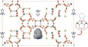 Crystal structure of the Hierarchical MESO-Microporous Zeolite ITQ-43 including the template molecule inside the pores solved by delta recycling method using automated diffraction tomography (ADT) data
Application of delta recycling to electron ADT data from inorganic crystalline nanovolumes, J. Rius, E. Mugnaioli, O. Vallcorba and U. Kolb,
Crystal structure of the Hierarchical MESO-Microporous Zeolite ITQ-43 including the template molecule inside the pores solved by delta recycling method using automated diffraction tomography (ADT) data
Application of delta recycling to electron ADT data from inorganic crystalline nanovolumes, J. Rius, E. Mugnaioli, O. Vallcorba and U. Kolb,Acta Cryst A, 69 396-407 (2013) - highlighted.
Solution Synthesis of a new thermoelectric Zn1+xSb nanophase and its structure determination using automated electron diffraction tomography, Birkel, E. Mugnaioli, T. Gorelik, M. Panthöfer, U. Kolb, W. Tremel, J.Am.Chem.Soc., 2010, 132(28), 9881-9889, DOI: 10.1021/ja1035122
Synthesis and Structure Determination of the Hierarchical MESO-Microporous Zeolite ITQ-43, Jiang, J.L. Jorda, J. Yu, L.A. Baumes, E. Mugnaioli, M.J. Diaz-Cabanas, U. Kolb, A. Corma, Science, 2011, 333(6046), 1131–1134, DOI: 10.1126/science.1208652
Ab Initio Structure Determination of Vaterite by Automated Electron Diffraction, E. Mugnaioli, I. Andrusenko, T. Schüler, N. Loges, R. Dinnebier, M. Panthöfer, W. Tremel, U. Kolb, Angewandte Chem.Int. Ed., 2012, 51(28), 7041-7045, DOI: 10.1002/anie.201200845
The Bi sulfates from the Alfenza Mine, Crodo, Italy: An Automatic Electron Diffraction Tomography (ADT) Study, G.C. Capitani, P. Gentile, T. Catelani, A. Lucotti, E. Mugnaioli, R. Branscheid, U. Kolb, American Mineralogist, 2014, 99(2-3) 500-510, DOI: 10.2138/am.2014.4446